Why Do Liver Transplants Require So Much Blood?

Liver transplants. A person's second chance at life. A truly amazing feat of medicine. And an occasional bane for Blood Bankers. Even the smoothest of surgeries generally take SOME blood. Over the last few decades blood usage has decreased dramatically overall due  to better techniques and advances in medicine, but the risk of bleeding still exists. Why?

The liver. Can't live without it!

Liver Transplant Bleeding


High Vascularity: The liver is one of the most blood-rich organs in the body, containing about 10% of the body’s blood supply at any given time.The liver's substantial blood volume, sourced from both the hepatic artery and the portal vein, underscores its high vascularity, elevating the complexity and potential bloodiness of liver-related surgical procedures. This dual blood supply system not only highlights the liver's critical role in bodily functions but also signifies the importance of meticulous surgical precision during liver transplants to manage and minimize blood loss effectively.

Complex Vascular Structures: The liver's vascular architecture, including major vessels entering and leaving the liver, makes surgical intervention complex. Precisely connecting these vessels during a transplant is critical to ensure the liver's blood supply in the recipient, and this process can lead to significant blood loss.

Disease State: Many patients requiring a liver transplant have underlying liver diseases such as cirrhosis, which can lead to altered blood clotting mechanisms and increased risk of bleeding. Moreover, liver disease often causes splenomegaly (enlarged spleen), which further complicates the surgery and increases the risk of bleeding.

Technical Challenges: The surgery involves removing the diseased liver and replacing it with a healthy one, connecting the blood vessels and bile ducts precisely to ensure the new liver functions properly. This process is technically challenging and can be associated with significant blood loss, especially in cases where there are anatomical variations or complications.

Advancements in surgical techniques, better understanding of liver anatomy, and improvements in preoperative planning and postoperative care have helped reduce the blood loss associated with liver transplants over the years. This includes the use of cell saver technology during surgery to collect and reinfuse the patient's own blood, minimizing the need for blood transfusions.

Anhepatic Phase 

The anhepatic phase during a liver transplant is a critical and unique period where the patient's diseased liver has been removed, and the new liver has not yet been fully connected and made functional. This phase is particularly challenging for several reasons:

No Liver Function: Since the liver performs many vital functions, including detoxification, protein synthesis, and production of clotting factors, its absence means these processes are temporarily halted. This can lead to an accumulation of toxins and a disruption in the body's metabolic balance.

Blood Clotting Issues: The liver is integral in producing clotting factors, so during the anhepatic phase, the patient is at an increased risk of bleeding due to reduced clotting factor production. This is compounded by the already complex vascular nature of the surgery and the potential for significant blood loss.

Hemodynamic Instability: The liver plays a role in regulating blood volume and pressure. Its removal can lead to hemodynamic instability, characterized by fluctuations in blood pressure and cardiac output. Managing the patient's fluid balance and circulation becomes more challenging during this phase.

Risk of Metabolic Complications: The liver's role in metabolism means its absence can lead to issues such as hypoglycemia. Monitoring and managing the patient's blood sugar and metabolic state are crucial during this time.

Due to these challenges, the anhepatic phase is a period of significant physiological stress for the patient. Anesthesiologists and surgical teams closely monitor vital signs, blood loss, and fluid balance. They may use various techniques and medications to support the patient's body until the new liver can be connected and begin functioning, thus ending the anhepatic phase. The shorter this phase can be kept, the better it is for the patient's overall outcome, as it reduces the time the body is without the critical functions provided by the liver.

Platelet Usage 

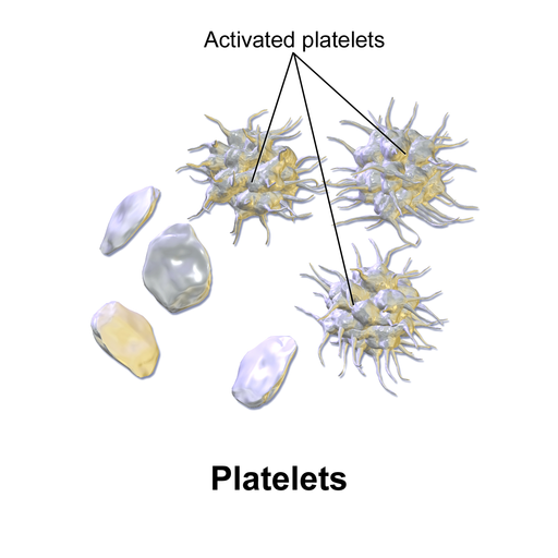
During liver transplant surgery, the consumption of a high number of platelets can be attributed to several factors, each emphasizing the complex interplay between liver function, surgical intervention, and the body's hemostatic processes:

Massive Blood Loss and Transfusions: Liver transplant surgeries often involve significant blood loss, requiring large volumes of blood transfusions. Transfused blood, especially when stored, may have reduced platelet function or count, necessitating additional platelet transfusions to maintain adequate hemostasis.

Dysfunctional Platelets: Patients with liver diseases, particularly those severe enough to require a transplant, often have dysfunctional platelets despite a normal or elevated platelet count. The liver plays a crucial role in producing coagulation factors and regulating platelet production. Liver disease can lead to thrombocytopenia (low platelet count) and qualitative platelet defects, making it necessary to transfuse platelets during surgery to correct these issues.

Activation of Coagulation System: The surgical stress and the extensive tissue manipulation during liver transplantation can activate the body's coagulation system. This activation can lead to the consumption of clotting factors and platelets, further necessitating the transfusion of platelets to maintain hemostasis.

Hypothermia: Liver transplants are long procedures, during which patients may become hypothermic. Hypothermia can impair platelet function, contributing to the need for platelet transfusions.

The Anhepatic Phase: As mentioned previously, during the anhepatic phase of the transplant, the patient's liver is not functioning, leading to an accumulation of toxins and further impairment of coagulation factors and platelet function. This phase can significantly contribute to the need for platelet transfusions.

The last liver transplant that came through our hospital took 16! SIXTEEN! Units of single donor apheresis platelets! Crazy. 

Blood Availability and Crossmatch 

The surgeon or anesthesia team will usually place blood orders ahead of time so Blood Bank can have blood products available for the surgery. Most of the time they request quite a bit of product up front, because of the 'bloody' nature of the surgeries. A previous surgeon would have a Liver Transplant Blood Package order of 8 RBCs, 8 FFP, and 4 platelets on hold. The new surgeon that took over wanted 4 whole blood, 10 RBCs, 10 FFP, and platelets/cryo as they go. Pretty wild. We basically had to change our entire policy for the surgeon, as our whole blood units were initially only intended and validated for use on adult male traumas in the ER.

Anti-Jk3 -- The Other Kidd

No not a child! Kidd!
Kidd Genesis --- Jk-Null Phenotype and Anti-Jk3

The discovery of the Jk-null phenotype, denoted as JK(a-b-), traces back to 1959 when a woman developed jaundice following a blood transfusion. Her serum contained an unusual antibody that recognized both Jka and Jkb antigens. This antibody was termed anti-Jk3.

Genetics and Population Prevalence

The Jk-null phenotype is exceptionally rare in most populations but somewhat more common in Polynesians, with a frequency of 0.9%. It is inherited as a recessive trait, implying that both parents must carry the mutated gene for a child to manifest this phenotype. Various mutations have been identified, with splice site mutations causing loss of exon 6 in Polynesians, and even different genetic mutations seen in the Finnish population.

Clinical Implications of Anti-Jk3

Hemolytic Disease of the Newborn (HDN)

Once immunized, individuals with the Jk-null phenotype can produce anti-Jk3 antibodies, which can result in HDN in subsequent pregnancies if the baby carries either Jka or Jkb antigen.

Hemolytic Transfusion Reactions

Anti-Jk3 can cause severe hemolytic transfusion reactions, both immediate and delayed, as it reacts against both Jka and Jkb. This makes finding compatible blood for transfusion exceedingly difficult for these individuals, as they can only receive JK(a-b-) blood.

The Physiology of Kidd Glycoprotein

The SLC14A1 gene encodes the Kidd glycoprotein, a significant urea transporter in red blood cells (RBCs). This transporter helps maintain osmotic stability by facilitating urea's rapid movement in and out of RBCs. In the kidney, this protein concentrates urine by allowing the medulla to maintain high urea concentrations.

In individuals with the Jk-null phenotype, urea transport across the RBC membrane is about 1,000 times slower than in those with typical RBCs. Despite this, the absence of the Kidd glycoprotein does not seem to have a substantial disease association. These RBCs maintain normal shape and lifespan, and the individuals show no other significant health issues besides an inability to maximally concentrate urine.

The Role of 2M Urea in Anti-Jk3 Characterization

In the domain of immunohematology, 2M urea serves as a powerful tool for antibody identification, particularly for Kidd antibodies. The Kidd blood group system antibodies, including anti-Jk3, are known for their sensitivity to 2M urea, which helps in their identification and characterization.

  1. Antibody Discrimination: Treatment of reagent RBCs with 2M urea can help distinguish Kidd antibodies from other blood group antibodies. If a suspected anti-Jk3 antibody retains its reactivity after 2M urea treatment, it is likely not a Kidd antibody. Conversely, if the reactivity is diminished or abolished, it suggests that the antibody is indeed anti-Jk3.

  2. Continuous Monitoring: For patients known to possess anti-Jk3, periodic reactivity checks can be performed using 2M urea. This is particularly important when planning for transfusions or transplants, as the presence of anti-Jk3 significantly narrows the range of compatible donor blood.

  3. Research Utility: In a more advanced research setting, 2M urea could be employed to study the thermodynamics and kinetics of anti-Jk3 binding to Jka and Jkb antigens. Such studies could offer insights into the mechanisms underlying the formation and specificity of anti-Jk3.

Anti-Jk3 is a clinically significant but rare antibody that primarily occurs in individuals with the Jk-null phenotype. Its presence poses challenges in transfusion medicine and maternal-fetal medicine. 2M urea plays a crucial role in the antibody's identification and ongoing monitoring. While the Kidd glycoprotein has a vital role in urea transport, its absence in Jk-null individuals does not cause disease, but it does lead to a significantly increased risk of hemolytic reactions in the context of transfusions and pregnancy. Therefore, understanding the complexities surrounding anti-Jk3 is crucial for better clinical management and future research.

Anti-PP1Pk and Pregnancy

Pregnacy and Anti-PP1Pk. Not a good mix
In women with a rare p phenotype, which lacks P, P1, and Pk red cell antigens, there is a presence of naturally occurring anti-PP1Pk antibodies, previously referred to as Anti-Tja. These antibodies have been closely associated with recurrent miscarriages, particularly in the first half of pregnancy. The p phenotype is exceedingly rare, with an estimated global prevalence of around 5.8 in one million people.

When a woman with this phenotype becomes pregnant, it's vital to manage the titers of the anti-PP1Pk antibodies, as they can pose risks to the pregnancy. In practice, this often involves treatments like plasma exchange therapy or double-filtration plasmapheresis. These treatments aim to reduce the concentration of these antibodies to safer levels (between 1:16 and 1:32) as a way to mitigate the risks.

However, the necessity of these aggressive treatments can depend on the woman's specific medical situation. For instance, some cases have shown that when these antibody titers are naturally low and well-monitored, it might be possible to manage the pregnancy without resorting to treatments like plasmapheresis. Monitoring usually involves bi-weekly checks of antibody titers.

In addition to managing the antibody levels, some patients are also put on medications like prednisolone and low-molecular-weight heparin (LMWH). While the exact role and effectiveness of these medications in such cases are still under debate, they are known for their anti-inflammatory and immunosuppressive properties.

The P, P1, and Pk antigens and the associated antibodies are part of broader blood group systems involving different glycosyltransferases necessary for their synthesis. The absence of these antigens, which gives rise to the p phenotype, is a result of inactivating mutations in the A4GALT1 gene located in chromosome 22q13.2. This gene is responsible for synthesizing 4-α-galactosyltransferase, the enzyme necessary for producing Pk, P, and P1 antigens.

Interestingly, these antibodies can exist in various forms, including regular IgM, irregular or regular IgG types, or a combination. The cytotoxic effects seem primarily to belong to the IgG3 subclass. These antibodies can cross the placental barrier and are known to activate complement, contributing to antibody-mediated cytotoxicity.

Identification of PP1Pk in hospital blood banks 

The presence of anti-PP1Pk antibodies would indeed yield results that could resemble panagglutination because every cell in the panel would likely test positive. This is due to the high prevalence of P, P1, and Pk antigens in the general population, making almost all donor cells susceptible to agglutination by the anti-PP1Pk antibodies. Therefore, distinguishing anti-PP1Pk from other causes of panagglutination would require specialized testing, often involving the use of cells specifically lacking these antigens or additional serological techniques. Hospital systems would be wise to send reference testing on patients, especially women of childbearing age, to determine the etiology of reactivity. 

Finding PP1Pk RBCs

Finding compatible blood products for individuals with anti-PP1Pk antibodies is an incredibly challenging task given the rarity of donors lacking P, P1, and Pk antigens. Standard blood banks are not equipped to handle this level of specificity in their inventories.

In these cases, medical professionals often have to resort to specialized approaches. The Rare Donor Program may be employed to find a compatible donor, although the rarity of such donors makes this a challenging and time-consuming endeavor. Even within this program, finding a matching donor can be like finding a needle in a haystack, given that these antigens are present in more than 99.9% of the general population.

An alternative approach could be the use of frozen blood products, assuming they have been previously identified and stored for this specific purpose. However, this is often not a guarantee, and there may be logistical and stability issues associated with using frozen products.

Due to these extreme difficulties, patients are sometimes encouraged to recruit potential donors from within their familial or community circles, although this too can be a long shot. If found, such donors can be invaluable, not only for the individual patient in need but also for adding to the database of rare donors.

Given these challenges, preemptive steps like autologous blood donation (where the patient donates blood for their own future use) may be considered, especially if a planned surgical procedure or childbirth is anticipated.

I actually had a case of a young pregnant woman with an Anti-PP1Pk. We notified our Blood Center several weeks prior to her scheduled delivery date that we would need two units of blood on hold as agreed upon by OB and Pathology. Come time for delivery, we had nothing on our shelves. They're search came up with nothing all that time, but thankfully mom delivered, with no complications! I shudder to think what would happen if things didn't go as planned!

Parvovirus B19 Receptor and PP1PK System

Parvovirus B19 primarily infects red blood cells by binding to the P antigen, which serves as its cellular receptor. The P antigen is a globoside and is essentially the foundation molecule for the other antigens (P1 and Pk) in this system. Individuals lacking the P antigen (rare but possible) are resistant to parvovirus B19 infection, which is known for causing diseases like erythema infectiosum (fifth disease) in children and temporary aplastic crises in adults with chronic hemolytic anemia.


Platelet Refractoriness

Platelet refractoriness is a significant challenge in transfusion medicine, referring to the failure of a patient to achieve the expected increase in platelet count following a platelet transfusion. This phenomenon has both immune and non-immune causes and can complicate the clinical management of patients, particularly those with hematologic malignancies or undergoing stem cell transplantation.

Causes of Platelet Refractoriness

Immune-Mediated Causes

1. Alloimmunization to HLA Class I Antigens
Platelets can be troublesome!

Human Leukocyte Antigens (HLA) are complex molecules that serve as the primary determinants for tissue compatibility in humans. Class I HLA molecules are present on almost all nucleated cells, including platelets. In the setting of platelet transfusions, the recipient's immune system can recognize these HLA antigens as foreign if they are not already present in the patient, leading to an immune response.

Mechanism of Alloimmunization

  • When a patient receives platelet transfusions from different donors, each transfusion episode carries a risk of introducing platelets with foreign HLA antigens. The immune system, recognizing these as non-self, can produce antibodies specific to these HLA antigens. Once alloimmunized, the recipient's plasma will contain these antibodies, which are primed to neutralize any subsequent transfusions containing platelets with the same or similar HLA antigens.

    Consequence

    The development of HLA antibodies leads to rapid clearance of transfused platelets from the circulation, resulting in suboptimal or negligible increases in platelet counts after transfusion. This means that even though the patient is receiving platelets, the expected improvement in clotting capability is not achieved, putting the patient at risk of bleeding complications.

    Diagnostic Strategies

    HLA Antibody Screening: This involves testing the patient’s serum for the presence of HLA antibodies. High-resolution assays like Luminex-based techniques can identify specific HLA antibodies, giving a more targeted approach to donor selection.

    Crossmatching: Before a transfusion, the donor's platelets can be crossmatched with the recipient's serum to test for compatibility.

Management Options

HLA-Matched Platelets: The ideal strategy is to transfuse platelets that are HLA-matched to the recipient. This minimizes the risk of antibody-mediated platelet destruction.

Cross-Matched Platelets: If an exact HLA match is not available, platelets that have been cross-matched to be compatible with the recipient can also be an option, although not commonly performed.

HLA-Desensitization: In extreme cases, immunosuppressive medications or plasmapheresis can be used to reduce the levels of HLA antibodies, although these options have their own set of risks and limitations.

Challenges in Clinical Practice

Donor Pool: Finding a suitable donor is often challenging and becomes progressively harder with each transfusion episode that leads to additional alloimmunization.

Logistical Constraints: HLA-matched platelets may not always be readily available and require coordination between different blood banks and registries.

Ethnic Variability: HLA types can vary between different ethnic groups, complicating matching in ethnically diverse populations.

Cost: High-resolution HLA typing and antibody screening tests are expensive and may not be feasible in all healthcare settings.

HLA MATCHED VS HLA AVOIDANCE 

HLA-Matched Platelets

Definition:

In this approach, the aim is to find a donor whose HLA antigens closely match those of the recipient. This reduces the chance of an immune response against the transfused platelets, leading to a more effective transfusion.

Methodology:

  • Comprehensive HLA typing is performed for both the recipient and potential donors.
  • Sophisticated matching algorithms may be used to find the closest possible match based on the HLA typing data.

Advantages:

  • Lower risk of transfusion reactions and refractoriness, as the risk of antigen-antibody interaction is minimized.
  • Can be highly effective in patients with known HLA antibodies, as the transfused platelets are less likely to be targeted for destruction.

Disadvantages:

  • Finding a close HLA match can be time-consuming and may not always be possible, especially in ethnically diverse populations.
  • More expensive due to the costs of HLA typing and the specialized handling and coordination required.

HLA Avoidance (also known as Antigen-Negative or HLA-Compatible Platelets)

Definition:

In this approach, the aim is not necessarily to match all HLA antigens between donor and recipient but to avoid those specific HLA antigens against which the recipient has developed antibodies.

Methodology:

  • The recipient is tested for HLA antibodies to identify the specific antigens that should be avoided.
  • Platelet units from donors lacking these particular antigens are then selected for transfusion, even if they are not a complete HLA match.

Advantages:

  • Faster and often easier to implement than finding a perfect HLA match, as you only need to avoid specific antigens rather than match all of them.
  • May be more readily available and cost-effective as compared to HLA-matched platelets.

Disadvantages:

  • While the risk of reaction is reduced, it is not as low as with HLA-matched platelets.
  • May still result in alloimmunization against other HLA antigens not previously sensitized against, as it's not a complete match.

____________________________________________________________________________________

2. Platelet-specific Alloantibodies: 

Human Platelet Antigens (HPA) are specific glycoproteins found on the membrane of platelets. Unlike HLA antigens, which are ubiquitously found on all nucleated cells, HPAs are specific to platelets. They serve as the scaffold for the binding of platelets to other cells and elements within the blood and are crucial for platelet function.

Mechanism of Alloimmunization

When a patient receives platelets from a donor with different HPAs, the recipient’s immune system can recognize these antigens as foreign and mount an antibody-mediated response against them. Like with HLA alloimmunization, the production of these antibodies is a learned response that sensitizes the immune system against future exposures to the same or similar HPAs.

Consequence

Once alloimmunized against a specific HPA, any subsequent transfusion containing platelets expressing this HPA will be targeted by the recipient's immune system. The result is rapid clearance of these transfused platelets, leading to inadequate increases in platelet count and a persistent risk of bleeding complications.

Diagnostic Strategies

HPA Antibody Screening: Similar to HLA antibody screening, this involves testing the patient’s serum for antibodies against specific HPAs. Techniques such as enzyme-linked immunosorbent assays (ELISAs) can be used for this purpose.

Platelet Crossmatching: This involves incubating donor platelets with the recipient's serum to check for compatibility, although this is more commonly done for HLA rather than HPA matching.

Management Options

HPA-Matched Platelets: These are platelets from a donor with matching or compatible HPAs to those of the recipient. Such matches are typically rarer than HLA-matches but can be highly effective in preventing refractoriness.

Immunosuppression: In extreme cases, immunosuppressive therapies may be considered to reduce antibody levels temporarily, but this comes with its own set of risks, including increased susceptibility to infections.

Challenges in Clinical Practice

Limited Awareness and Testing: HPA alloimmunization is not as well-known or as routinely tested for as HLA alloimmunization, which can result in underdiagnosis.

Donor Pool: Finding HPA-matched donors is often even more difficult than finding HLA-matched donors, particularly because routine platelet donors are not typically typed for HPAs.

Economic Considerations: Specialized tests for HPA antibodies and HPA-matched platelets can be costly and may not be available in all healthcare settings.

____________________________________________________________________________________

Non-Immune Platelet Refractory Causes

Fever and Sepsis

Mechanism: Elevated body temperature and systemic infections can lead to increased platelet consumption and turnover. In sepsis, there's often disseminated activation of the clotting cascade, which consumes platelets faster than usual.

Clinical Implication: A patient with fever or sepsis may have a lower post-transfusion platelet count increment, not because the transfused platelets are being destroyed by antibodies, but because they're being used up rapidly.

Drugs

Amphotericin B: An antifungal medication known to cause various side effects, including platelet aggregation, which might reduce the efficacy of platelet transfusion.

Heparin: An anticoagulant that can, in rare instances like heparin-induced thrombocytopenia (HIT), lead to platelet destruction.

Clinical Implication: Recognizing drug-induced thrombocytopenia is crucial, as the cessation of the offending drug can often reverse the platelet count decline.

DIC (Disseminated Intravascular Coagulation)

Mechanism: DIC is a disorder characterized by systemic activation of the blood clotting system. This leads to widespread formation of micro-clots in small blood vessels. As a result, platelets (and clotting factors) are consumed at an accelerated rate.

Clinical Implication: Patients with DIC may show poor increments in platelet counts after transfusion because of the rapid consumption of both endogenous and transfused platelets.

Splenomegaly

Mechanism: An enlarged spleen (splenomegaly) can sequester a larger portion of platelets than usual, reducing their circulating numbers.

Clinical Implication: Even after platelet transfusion, patients with significant splenomegaly might show suboptimal platelet count increments.

Graft-Versus-Host Disease (GVHD)

Mechanism: GVHD is a condition that can occur after stem cell or bone marrow transplantation, where donor cells attack the recipient's body. In the context of platelets, GVHD can contribute to bone marrow suppression, thus affecting platelet production.

Clinical Implication: Platelet refractoriness in GVHD isn't just about platelet destruction; it's also about reduced production. Addressing the underlying GVHD is crucial.

Bleeding

Mechanism: Active bleeding, especially in large amounts, can quickly consume the existing and transfused platelets.

Clinical Implication: In a bleeding patient, poor post-transfusion platelet count increments might be due to the immediate consumption of platelets at bleeding sites.

____________________________________________________________________________________

Clinical Evaluation and Diagnosis

The cornerstone of diagnosis is the calculated Corrected Count Increment (CCI) post-transfusion. A CCI lower than expected suggests refractoriness. Additional steps:

  • Blood Sample Analysis: To evaluate for alloantibodies against HLA and/or platelet-specific antigens.

  • Clinical Assessment: To rule out non-immune causes, like splenomegaly or sepsis.

_____________________________________________________________________________________

Platelet Refractory Management Strategies

 Immune Mediated:

  • HLA-Matched Platelets: Ideally from closely HLA-matched donors or family members.

  • Cross-matched Platelets: Selected based on the absence of the specific antigens against which the patient has antibodies.

  • Desensitization: Rarely used, this involves administering the triggering antigen in increasing doses to reduce the recipient's antibody response.

Non-Immune Mediated:

Address the underlying cause, be it infection, medication, or other conditions.

____________________________________________________________________________________

Platelet Refractoriness Prevention

Leukoreduction: Removing white blood cells from transfused blood products reduces the risk of alloimmunization.

Antigen Matching: For at-risk patients, consider HLA-matched or cross-matched platelets.

Hemovigilance: For at-risk patients, transfuse platelets only when clinically indicated. If patient is not bleeding or at risk of bleeding, consider whether holding off on transfusion is possible.

____________________________________________________________________________________

Future Directions and Innovations:

  • Genomic Matching: Advanced molecular techniques offer potential for better donor-recipient matching.

  • Platelet Growth Factors: Agents like thrombopoietin mimetics can stimulate platelet production, potentially reducing the need for transfusions.

  • Therapeutic Modalities: Immunomodulation or extracorporeal treatments, like plasmapheresis, may play roles in specific refractory cases.

Weak D and Partial D

The D antigen, a significant component of the Rh blood group system, plays a crucial role in transfusion medicine. While most individuals are categorized as either RhD-positive (presence of the D antigen) or RhD-negative (absence of the D antigen), there are more intricate phenotypes to consider: weak D and partial D. Understanding these nuanced categories is vital for ensuring safe blood transfusions and managing pregnancies. 

DNA -- check for Weak or Partial D!

Molecular genotyping is vital for Weak D and Partial D typing because of its precision and the clinical implications of accurate identification. The process involves extracting DNA, amplifying specific regions of the RHD gene, and then analyzing or sequencing these regions to determine the exact variant.

Molecular Basis and Phenotypic Differences

  • Weak D: Weak D is characterized by a reduced expression of normal D antigens on the red blood cell (RBC) surface. This could result from genetic mutations leading to changes in the RhD protein's structure, affecting its external display on the RBC. Traditional serologic testing might classify these individuals as D-negative, but more sensitive tests will detect the D antigen's presence.

  • Partial D: Unlike weak D, which is about the decreased expression of normal D antigens, partial D results from structural variations of the D antigen. Individuals with partial D express all the epitopes of the D antigen, but one or more are altered. This alteration might cause them to produce anti-D antibodies if exposed to normal D antigen, posing a transfusion risk.

Clinical Implications in Transfusion Medicine

  • Blood Transfusions: People with weak D are typically safe to receive D-positive blood without the risk of alloimmunization, as their body recognizes the D antigen, albeit in lower amounts. However, for individuals with partial D, there's a potential risk when receiving D-positive blood since they might produce anti-D antibodies against the epitopes they lack.

  • Pregnancy and Hemolytic Disease of the Newborn (HDN): RhD incompatibility between a mother and her fetus can lead to HDN. Typically, RhD-negative mothers are at risk of developing anti-D antibodies when carrying an RhD-positive fetus. For mothers with weak D phenotypes, the risk of alloimmunization is minimal. However, for partial D mothers, the situation is complex. Depending on the specific epitopes they lack, they may still be at risk for developing anti-D antibodies.

Diagnostic Challenges and Recommendations

Detecting weak and partial D phenotypes requires advanced serological testing. Standard agglutination tests might miss weak D expressions, leading to incorrect classifications. Molecular genotyping can provide a more precise identification of weak D and partial D phenotypes.

For transfusion purposes, it might be simpler and more conservative to consider individuals with weak or partial D as D-negative, especially if detailed phenotypic or genotypic analysis isn't feasible. However, this approach can lead to unnecessary administration of Rh immunoglobulin (RhIg) in pregnancy and limit the availability of D-negative blood, which is rarer and more precious.

Strategies for Management

  • Blood Transfusions: To prevent alloimmunization, individuals with partial D phenotypes should ideally receive D-negative blood. In emergencies, when D-negative blood isn't available, RhIg can be administered as a preventive measure.

  • Pregnancy: All pregnant women should undergo RhD typing. For those with weak D phenotypes, RhIg prophylaxis might not be necessary. However, those with partial D phenotypes should be managed as RhD-negative, with RhIg administration to prevent potential alloimmunization. 

    Weak D Types:

    The term "Weak D" is used to describe red blood cells (RBCs) that exhibit a weakened expression of the D antigen but produce all epitopes of the RhD antigen. Over 150 Weak D types have been described, but the common ones are:

    1. Weak D Type 1, 2, and 3: These are the most frequent types in Caucasians. From a clinical standpoint, individuals with these types are not at risk for D alloimmunization, even upon exposure to D-positive blood.

    2. Weak D Type 4: This group consists of multiple subtypes (e.g., Type 4.0, 4.1, 4.2, etc.). Some subtypes can produce an anti-D response when exposed to D-positive RBCs, especially the Type 4.2 variant. RhIg prophylaxis is often indicated in this case.

    3. Other Weak D Types: Many other types exist, with varying clinical significance. Their potential to produce an anti-D response upon exposure to D-positive RBCs varies.

    Partial D Variants:

    Partial D phenotypes arise due to genetic mutations leading to the production of an altered D antigen. The clinical risk associated with Partial D phenotypes pertains mainly to potential alloimmunization upon exposure to regular D-positive RBCs.

    DIVa/DIVb

    DIIIa, DIIIb, and DIIIc

    DVa and DVb 

    D^U (or DAU): A complex category, which includes numerous subtypes like DAU-0, DAU-1, DAU-2, etc. Their clinical relevance varies, but some DAU types can result in alloimmunization upon exposure to standard D-positive blood.

     DVI - The DVI variant represents an epitopic modification within the D antigen complex, resulting in the absence of certain epitopes that are present in a regular D antigen. DVI arises due to the presence of specific alleles of the RHD gene. The molecular characterization of DVI shows hybrid RHD-CE-D gene structures.

    And many more!!

    Management:

    1. Weak D: Most guidelines now suggest that individuals with Weak D Type 1, 2, or 3 can safely receive D-positive blood without the risk of alloimmunization. For other Weak D types, the decision should be made on a case-by-case basis, considering potential risks.

    2. Partial D: Due to the risk of alloimmunization, individuals with known Partial D phenotypes should receive D-negative blood, especially when the specific subtype's risk is not well-defined.

    3. Pregnancy: Women with Weak D types not associated with alloimmunization (e.g., Type 1, 2, 3) do not typically require RhIg prophylaxis. However, those with certain Partial D phenotypes or uncharacterized Weak D types may benefit from a more conservative approach, including RhIg administration and closer monitoring.


Subgroups of (Blood Type) B?

This post brought to you by...the Letter B!
Yes, they do exist! We are generally much more familiar with subgroups of A, especially in patients who have made an Anti-A1, but subgroups of B do exist in patient populations. They are rare, but this lack of prevalence can likely be partially explained by Blood group B having a much lower prevalence than A to begin with. 

Here's a breakdown of the B subgroups:

B3 Subgroup

Antigenic Expression:
  • The B3 subgroup presents with a weaker expression of the B antigen when compared to the regular B blood group. This diminished strength is often the result of changes in the sugar structures that form the basis of the B antigen.
Mixed-Field Agglutination:
  • B3 cells often display a phenomenon called mixed-field agglutination when tested with anti-B serums. Mixed-field agglutination refers to the simultaneous presence of agglutinated and non-agglutinated red cells in the same sample. This can occur if only a fraction of the RBCs in the sample express the B antigen, leading to partial clumping.
Genetic Inheritance:
  • The B3 phenotype appears to have an autosomal dominant pattern of inheritance. It's inherited in families, and if one parent carries the gene, there's a 50% chance that their offspring will also express this subgroup.
Differentiation from Acquired B Antigen:
  • Acquired B antigen often arises due to external factors, typically associated with diseases affecting the gastrointestinal tract. The enzymes produced by certain bacteria can modify the A antigen to resemble B antigen, leading to the "acquired B" phenotype.
  • Differentiating between B3 and acquired B is essential for several reasons:
    • Transfusion Reactions: Patients with acquired B can experience transfusion reactions if given blood from true B or AB donors. Knowing the difference helps guide appropriate blood product selection.
    • Organ Transplantation: Similar to transfusions, ensuring the correct blood type and its nuances are critical for organ transplants.
    • Diagnostic Significance: Acquired B antigen can be an indicator of an underlying disease. Recognizing this can be a diagnostic clue, especially in patients with unexplained anemia or those who have a history suggestive of gastrointestinal disease.

Further Considerations:

  1. Testing: Enhanced testing methodologies, including the use of different antisera or molecular techniques, may help clarify ambiguous results.
  2. Clinical Implications: While B3 is primarily of academic interest, its recognition can prevent potential transfusion-related complications. Blood bank specialists must be aware of its existence and its differentiation from the more clinically significant acquired B phenotype.

In summary, the B3 subgroup, while rare, represents a fascinating intersection of genetics, biochemistry, and clinical medicine within the realm of immunohematology.


Bx (Bend) Subgroup

Antigenic Expression:
  • The Bx subgroup, similar to B3, displays a weaker expression of the B antigen on the surface of red blood cells. However, what distinguishes Bx (Bend) cells from typical B cells is their unique reactivity pattern.
Reactivity with Anti-B:
  • The Bx cells demonstrate weak agglutination (clumping) when reacted with anti-B sera. This weak agglutination can sometimes be misinterpreted, leading to potential misclassification of the blood type, especially if other subgroups or acquired conditions aren't considered.
Temperature-Dependent Reactivity:
  • One of the defining features of the Bx (Bend) subgroup is its enhanced reactivity at colder temperatures, typically around 4°C. When Bx cells are tested at this lower temperature, the agglutination reaction with anti-B sera is stronger.
  • The temperature-dependent reactivity suggests specific structural or conformational changes in the B antigen that favor binding to the anti-B antibody at lower temperatures.
Clinical Implications:
  • Blood Transfusion: Recognizing the Bx (Bend) subgroup is critical for blood banks, as misclassification can lead to transfusion reactions. If a person with the Bx phenotype is mistyped as group O and then receives B or AB blood, it could cause a reaction.
  • Laboratory Practices: Because of its temperature-dependent reactivity, blood banks must be vigilant when performing tests, especially if the reactions are carried out at different temperatures. Moreover, if a blood bank specialist encounters unexpected weak B reactions, the Bx (Bend) subgroup should be considered, and additional tests at various temperatures may be conducted.
  • Genetics and Biochemistry: While the Bx (Bend) subgroup has been defined serologically, the genetic and biochemical underpinnings are not as well-understood as the main blood group types. There may be genetic mutations or alterations in the enzymes involved in synthesizing the B antigen, leading to the distinct Bx phenotype.

Bm (B modified) Subgroup

Antigenic Expression:

  • The Bm subgroup carries a modified or variant form of the B antigen on the surface of red blood cells. This modification leads to a differential reactivity when exposed to certain anti-B reagents.

Reactivity with Anti-B:

  • Bm cells typically show weaker reactions or sometimes even negative reactions with specific monoclonal anti-B reagents. This can create confusion in blood typing, potentially leading to misclassification.
  • The reactivity pattern varies depending on the origin of the anti-B reagent used, especially between monoclonal and polyclonal sources. Polyclonal anti-B sera, derived from multiple immune cells, might still show agglutination with Bm cells, while certain monoclonal reagents, originating from a single immune cell clone, might not.

Geographical and Ethnic Distribution:

  • The Bm phenotype has been found in various populations worldwide but remains relatively rare. Its presence in both African and Caucasian populations suggests that it's not limited to a specific ethnic group, though its prevalence might vary across different communities.
  • Genetics and Biochemistry: The specific genetic and biochemical basis for the Bm phenotype is not entirely clear. However, it is believed to arise from genetic mutations or alterations in the enzymes responsible for B antigen synthesis, leading to a modified structure.

Bel Subgroup

Antigenic Expression:

  • Individuals with the Bel subgroup have red blood cells that express an extremely low amount of the B antigen—so low that it's nearly indistinguishable from an O blood type using conventional serological methods.
  • Because of this minute expression, routine ABO typing may detect these cells as type O, leading to potential misclassification.

Anti-B Production:

  • Despite the almost non-existent B antigen expression, individuals with the Bel phenotype can produce anti-B antibodies. The production of this antibody suggests that the level of B antigen present on their cells is insufficient to induce tolerance.
  • The anti-B produced by Bel individuals is not benign. If a Bel individual receives blood from a B or AB donor, this anti-B can target and destroy the transfused red blood cells, leading to a hemolytic transfusion reaction—a serious and potentially life-threatening adverse event.

Clinical Implications:

  • Blood Transfusion: Given the potential for Bel individuals to produce anti-B, it's paramount to identify this subgroup accurately. Misidentification could lead to providing B or AB blood to a Bel recipient, which would trigger a hemolytic reaction.
  • Laboratory Challenges: The primary challenge is the potential misclassification of Bel as O due to the almost non-existent B antigen expression. Advanced serological methods, such as adsorption and elution techniques, or molecular tests may be required to detect and confirm the presence of the Bel phenotype.
  • Pregnancy and Hemolytic Disease of the Newborn (HDN): While the focus is often on transfusion, it's also essential to consider potential complications in pregnancy. If a Bel mother is pregnant with a fetus expressing the B antigen, there's a risk, albeit low, for HDN.

Historical Context:

  • The name "Bel" is derived from the initial patient's name in whom this phenotype was first identified. Over time, as with many blood group anomalies, the name has been used to represent the entire subgroup.

Acquired B Blood Group Phenotype

Nature of Change:

  • As the name suggests, this alteration in blood group is not inherited. Instead, it arises due to external factors during an individual's lifetime.
  • The phenomenon involves a modification of the A antigen on the red cell surface, causing it to mimic the B antigen.

Mechanism:

  • The transformation of the A antigen to resemble the B antigen is primarily attributed to the action of bacterial enzymes. Certain bacteria, especially those flourishing in gastrointestinal conditions, produce enzymes known as deacetylases.
  • These microbial deacetylase enzymes remove acetyl groups from the A antigen, altering its structure. The modified A antigen then mimics the B antigen in serological reactions.

Clinical Implications:

  • Blood Typing Discrepancies: The most immediate consequence of the Acquired B phenomenon is a discrepancy in blood typing. An individual previously typed as group A may appear as group AB due to the presence of the modified A antigen that reacts with anti-B sera.
  • Transfusion Concerns: Given the altered blood typing results, there's a potential risk of transfusion errors. However, it's important to note that the acquired B antigen typically does not trigger an immune response. Thus, even if group B blood was transfused into a patient with Acquired B, hemolytic reactions are unlikely. Still, it's crucial to adhere to blood transfusion guidelines and provide group-specific or type O blood when in doubt.

Associated Conditions:

  • Acquired B is most frequently associated with gastrointestinal diseases, where the growth and activity of certain bacteria are enhanced. Common conditions include:
    • Colon cancer
    • Intestinal obstruction
    • Peptic ulcers
    • Other gastrointestinal malignancies
  • The association with gastrointestinal diseases, especially colon cancer, implies that the presence of Acquired B could serve as a diagnostic hint, suggesting the need for further gastrointestinal evaluation.

Resolution and Diagnosis:

  • Acquired B is generally a transient phenomenon. Once the underlying gastrointestinal condition is addressed or if the microbial flora is altered (e.g., due to antibiotics), the blood group typically reverts to its original type.
  • If Acquired B is suspected, repeating the blood group test after treating the individual with acid or enzymes can revert the modified A antigen back to its original state, thus confirming the diagnosis.

Differentiation from Genuine AB:

  • In order to differentiate Acquired B from a true AB blood type, a detailed patient history is crucial. Salivary blood grouping, which remains unaffected by the acquired B phenomenon, can also aid in distinguishing between the two.

In essence, Acquired B serves as a testament to the dynamic nature of the human body and its interactions with the microbial environment. Recognizing such anomalies and understanding their underlying mechanisms ensures patient safety and can also provide valuable diagnostic insights.

B(A) Subgroup of Blood Group B

Nature and Origin:

  • The B(A) phenotype exhibits characteristics of both B and A antigens on the red cell surface, but the A-like properties are weaker than those seen in a true A antigen.
  • The origin of the A-like quality in these B cells isn't entirely clear. It's believed that this phenotype arises due to the activity of an A transferase enzyme that is functioning at a reduced capacity. This enzyme adds specific sugars to the H antigen, turning it into an A antigen. However, in the case of B(A) subgroup, this enzyme's activity isn't as efficient as in regular A blood group individuals, leading to a weaker A expression.

Serological Characteristics:

  • Blood samples from individuals with B(A) typically react with anti-B and anti-A sera, but the reaction with anti-A is weaker. This can cause discrepancies in blood grouping.
  • Monoclonal anti-A reagents might not detect the weaker A antigen, but polyclonal anti-A reagents (which are generally more sensitive) may produce a reaction.

Clinical Implications:

  • Blood Typing Confusion: The most significant concern with B(A) is the potential for misclassification. If not detected and classified correctly, a B(A) individual could be mistyped as AB or B.
  • Transfusion Issues: A person with a B(A) phenotype can produce anti-A antibodies, even though they have A-like properties on their cells. This means they can potentially have a transfusion reaction if given blood from a true A or AB donor.

Identification:

  • Thorough serological tests, often employing a series of anti-A reagents and adsorption-elution techniques, can help in correctly identifying this subgroup. Adsorption techniques can remove the interfering antibodies, and elution can then be used to identify the eluted antibodies.
  • A detailed patient history is also beneficial. Some B(A) phenotypes are acquired due to underlying conditions, similar to the Acquired B phenomenon.

Epidemiology:

  • The prevalence of the B(A) subgroup varies among populations but is generally rare.
  • Some studies suggest a higher occurrence in certain Asian populations, though it's still a rare phenomenon.


Donath-Landsteiner Antibodies

For many, the intricacies of the human immune system remain an enigma, especially when it comes to rare autoimmune conditions. Today, we are diving deep into one such mysterious condition: Paroxysmal Cold Hemoglobinuria (PCH), and the unique antibody associated with it - the Donath-Landsteiner (D-L) antibody.

The Enigma of PCH and its Association with D-L Antibodies

Paroxysmal Cold Hemoglobinuria, or PCH, is an unusual autoimmune hemolytic anemia. In simpler terms, it's where the body's defense system, mistakenly, targets and destroys its own red blood cells (RBCs). What sets PCH apart from other anemias is its trigger: cold temperatures.

While PCH can strike any age group, children and young adults are particularly vulnerable. Historically linked to syphilis in adults, PCH today is more commonly seen following viral infections in children. This condition is typically transient in nature but can cause significant acute symptoms.

Donath-Landsteiner hemolytic anemia has been correlated with various viral infections, including:

  • The Epstein-Barr virus (commonly known as EBV).
  • Coxsackievirus type A9.
  • Adenovirus.
  • The Cytomegalovirus, abbreviated as CMV.
  • Parvovirus.
  • Varicella zoster virus, the causative agent of chickenpox.
  • Mumps.
  • Measles.

The main culprit behind this self-destructive process is the Donath-Landsteiner antibody. What's fascinating about this antibody is its specificity. Unlike other autoantibodies, D-L antibodies specifically target a carbohydrate antigen called the P antigen found on the surface of RBCs.

The Cold Activation Mechanism

D-L antibodies stand out in the realm of immunology due to their distinctive behavior. Unlike the typical IgM antibodies, which commonly react at cold temperatures, D-L antibodies are predominantly of the IgG class. Yet, they share the unique characteristic of reacting at cold temperatures, much like their IgM counterparts. This dual peculiarity—being an IgG that reacts to cold—makes them particularly notable in the field of blood bank and transfusion medicine.

D-L antibodies are uniquely temperature-sensitive. During exposure to cold, these antibodies bind to the P antigen on RBCs. As the blood rewarm, typically when it circulates back to the core of the body, the complement system - a component of the immune system - gets activated. This activation results in the destruction (hemolysis) of RBCs.

Symptoms can vary from mild fatigue to severe anemia, with dark or red urine indicating the presence of hemoglobin from the lysed RBCs. This condition can be alarming, especially if there's an extensive destruction of RBCs, leading to acute anemia.

The Diagnostic Challenge

Diagnosing PCH requires a high degree of clinical suspicion. The connection between cold exposure and the onset of symptoms is crucial for diagnosis. Standard blood tests may reveal anemia and evidence of hemolysis, such as elevated bilirubin, low haptoglobin, and increased lactate dehydrogenase (LDH).

However, the definitive diagnostic test is the Donath-Landsteiner test. This test exposes the patient's blood to cold temperatures, then warms it to body temperature to see if hemolysis occurs in the presence of complement. A positive test will confirm the presence of D-L antibodies and the diagnosis of PCH.

Treatment Strategies

Managing PCH revolves around avoiding triggers and treating acute episodes. Since cold exposure is a primary trigger, patients are often counseled to avoid cold environments and ensure their extremities are well-protected during chilly days.

Treatment during acute hemolytic episodes may involve blood transfusions to replenish the RBCs lost to hemolysis. Immunosuppressive medications like corticosteroids can also be used to temper the immune response. In rare, persistent cases, stronger immunosuppressive drugs or even a bone marrow transplant might be considered.