RhIG to an Rh positive Patient? The Medical Splenectomy

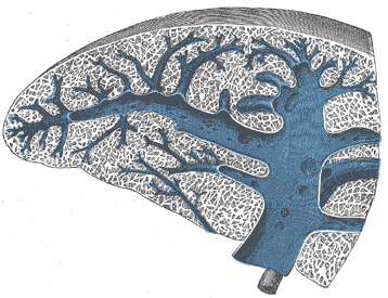
Spleen
 The nursing floor just requested RhIG on an Rh positive MALE? Why???

They likely want to perform a "medical splenectomy"!

Patients with ITP or idiopathic (or immune) thrombocytic purpura create an autoantibody towards antigens on platelets which subsequently marks the platelets for destruction and they are removed from circulation causing thrombocytopenia. Typically, physicians will try treatments such as steroids first, to dampen the immune systems response and see if platelet counts recover. If not, there are other options available. Very often physicians will spring for IVIg (Intravenous Immunoglobulin). The mechanism in which IVIg works in regards to platelets in ITP is not fully understood but it is postulated the immunoglobulins bind receptors on macrophages, leaving them unable to interact with platelets and target them for destruction. This inability to mark the platelets for destruction means more platelets remain in circulation. 

There is another option, however, and it is still considered a first-line therapy, much like IVIg is. RhIG! Yes, Rh immune globulin, the same stuff you've been giving Rh negative pregnant mothers all this time to prevent RhD alloimmunization. WinRho SDF and Rhophylac brands are approved for IV usage in ITP situations. RhoGAM (a specific brand of RhIG) is typically given only as an IM shot. RhoGAM can ultimately be used, but patients may need multiple shots, making WinRho and Rhophylac a better choice. 

To receive IV RhIG for ITP treatment, patients MUST be Rh positive, must still have a functional spleen, and should not be exhibiting signs of hemolytic anemia or DIC. Providing RhIG to Rh positive patients for treatment of ITP is known colloquially as performing a "medical splenectomy". This is because removing the spleen is a drastic measure to stop platelet sequestration and destruction, as it is where the platelets get sent for destruction once they are antibody targeted. In more medically scientific terms it is know as an Fc receptor blockade mechanism. 

This mechanism can be described as such:

The anti-D from the RhIG preparation attaches to the RhD positive red cells of the recipient. Essentially, the RhIG "opsonizes" the RhD red blood cells, targeting them for destruction. The RhD positive red cells are brought through the reticuloendothelial system via phagocytic cells like macrophages where they will be destroyed in the spleen. This massive increase in antibody marked cells, coupled with the fact there are far more antigen sites on a Red Blood Cell, meaning more antibody can attach, means that the Red Cells will generally preferentially get removed from circulation rather than the platelets, allowing the platelets to remain in circulation. 

Given this mechanism, it is not only possible, but likely that the patient can experience a level extravascular hemolysis leading to a mild decrease in Hemoglobin/Hematocrit levels. It's important to get a baseline, and continue to monitor these levels after administration. A drop of 1-2 grams of Hgb is possible over a few days post administration. This is also why it's important to ensure there aren't other hemolytic processes taking place beforehand (like hemolytic anemia). If a patients Hgb drops too much, it is thought that they should receive Rh negative blood for the time being, to lessen the amount of hemolysis taking place, ensuring the blood remains in circulation unscathed. 

Despite these effects, use of RhIG is often quicker to infuse than IVIG, less volume than IVIG, sometimes longer duration of action when compared to IVIG, cheaper than IVIG, and a much more limited donor exposure than IVIG. 

Have you ever seen RhIG used in such circumstances?
Emergent FFP for Angioedema?

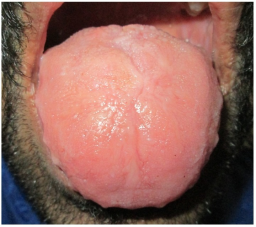
You get a call from an ER nurse that they new FFP emergently on a patient in the ER. The patient's tongue and face is swelling to the point of respiratory compromise. You don't have any blood type on file so you tell the nurse, and decide that it's so emergent that you'll have to thaw AB plasma and give it emergently. 

Macroglossia with crenations along the margins and loss of papillae on dorsum surface of the tongueWhat was happening to this patient? Why was this such an emergency? What is FFP going to do to help this patient in this situation?

Angiotensin Converting Enzyme (ACE)

 Millions of people in the world have diagnosed hypertension and may be prescribed an Angiotensin Converting Enzyme (ACE) inhibitors to control their elevated blood pressure. An uncommon side effect of ACE inhibitors includes angioedema, in which fluid buildup and swelling occurs underneath the skin in certain areas. Inhibiting Angiotensin Converting Enzyme can lead to a build up of an enzyme known as bradykinin, which is a potent vasodilator, but also leads to increased vascular and capillary permeabilty. This allows fluids to leave the space they should occupy and accumulate in other areas in which they shouldn't. This effect is known as ACE inhibitor-induced angioedema or ACEI-IAE.

ACEI-IAE typically presents as an emergency as airways and respiratory systems can quickly become compromised by excessive tongue, face, neck, etc., swelling. Initial treatment will include discontinuation of the drug, and potentially antihistamines, steroids, epinephrine, IVIG, etc. This may not work for all patients, however. 

How Does FFP Help with Angioedema?

FFP has been used successfully in patients who do not respond to other treatments. Some patients respond quite rapidly after FFP transfusion and regain respiratory control. The idea behind FFP administration is that FFP contains ACE, sometimes referred to as Kininase II, as it's an enzyme that can break down Bradykinin. This immediate addition of Kininase II from FFP transfusion helps to breakdown the excessive bradykinin that has accumulated due to ACE inhibitor use. This is why so many patients will see a very rapid improvement post FFP administration. 

Blocked D Phenomenon

Why would an Rh positive baby type as Rh negative?

Intrauterine transfusion? Sure that's possible. 

This specific scenario describes more of an immunologic basis for mistyping.

Enter...the Blocked D phenomenon. It is something very rarely seen in Blood Banks, especially these days with ever increasing sensitivity of reagents, but it can still happen. 

The Blocked D phenomenon may occur when a mother has created a strong IgG based allo-Anti-D and bears an Rh positive child. It is generally seen in fairly severe cases of HDFN. 

A sample experiencing the blocked D phenomenon will type as Rh negative, and essentially always have a positive IgG DAT. A subsequent elution would show the maternal Anti-D coating the Red Cells of the baby. The red cells type as Rh negative because maternal Anti-D is blanketing the RhD antigens on the Red Cells, blocking the REAGENT IgM Anti-D from agglutinating the cells. As a result, a false negative Rh testing occurs. With a positive IgG DAT, it would not be possible to perform a Weak D (Du) without treating the cells either, as Weak D testing is performed at the AHG (Coombs) phase. 

Cord Blood Evaluations generally arrive to the blood bank as a battery of Blood Type and Direct Coombs testing. This is why it is important to finish all testing prior to resulting. If the blood type was resulted before the DAT was completed, you may be inadvertently reporting erroneous results to the patient's Blood Bank file and chart. 

How to remedy this?

Generally you would need to perform an elution, such as an acid glycine elution, to remove the bound IgG from the neonates Red Cells. Once removed, and you can prove the antibody is removed by perform another DAT to make sure the IgG is now negative, you can retest the red cells for the D antigen using your monoclonal typing reagents. You may now perform a Weak D test as well, since the DAT is no longer IgG positive. 

How many of you have seen this phenomenon? While rare, it has real implications in the Blood Bank and for proper patient care. Not realizing the phenomenon taking place, can result in a delay of care. A newborn exhibiting this phenomenon may need to receive an exchange transfusion to remove their Red Cells and replenish with Rh negative cells until the maternal antibody is no longer reacting. If the phenomenon is not noticed at first, and the baby is resulted as Rh negative, it will change the clinical picture for the physician and make it more difficult for them to arrive at a diagnosis of HDFN and treat at a earlier point in time. 


Fat Embolism Syndrome in Sickle Cell Disease

Sickle cells
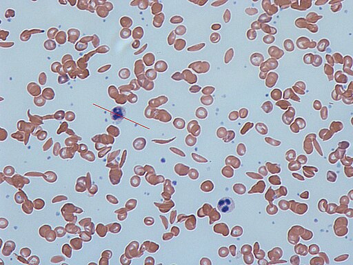
Sickle Cells on a peripheral blood smear

 Blood Banks are no stranger to patients with Sickle Cell Anemia. Many patients are chronically transfused, and many of us have been there for emergent Red Blood Cell exchanges due to acute chest, stroke, etc. What you might not have been a part of is a Sickle Cell patient experiencing Fat Embolism Syndrome. 

What is a Fat Embolism?

A fat embolism is exactly as it sounds, it's fat or globules of fat that for one reason or another enter the bloodstream circulation and act as a embolism (vasculature occlusion caused by a clot or similar). While not common, fat embolisms are usually seen in trauma with orthopedic / long bone fractures, such as tibia, fibula, femur, pelvis, etc. It can also be seen in patients with pancreatitis. 

Fat Embolism Syndrome in Sickle Cell patients

Certain populations of Sickle Cell patients may be at risk for Fat Embolisms as well. 

Fat Embolism Syndrome describes a situation in which Bone Marrow Necrosis occurs as a result of the patients Sickle Cell Disease. The necrosis is not fully understood but it's hypothesized that the microvasculature in the bone marrow may become occluded, leading to cell damage and death. As the Bone Marrow is necrosed, it is thought that this could lead to fat emboli being released into circulation, causing occlusion of vessels. Another theory proposes that fat globules released into circulation get broken down into toxic metabolites leading to a pro-inflammatory state, as evidenced by increased levels of CRP, cytokines, and free fatty acids in serum. These metabolites can be responsible for many of the symptoms seen in Fat Embolism Syndrome. 

Patients may have shortness of breath/respiratory distress, tachycardia, neurologic changes from confusion up to coma, petechial rash, pain, fever, hepatic damage (with resulting jaundice), decreased urine output, etc. 

Interestingly, patient's with more severe homozygous Sickle Cell Disease (HgbSS) are less likely to experience Fat Embolism Syndrome. Heterozygous Sickle Cell Disease such as HgbSC or HbS/ß-Thalassemia has a higher likelihood of exhibiting Fat Embolism Syndrome propensity. The thought behind this is that with heterozygous Sickle Cell Disease, patients tend to have a higher baseline hematocrit, and thus have a higher blood viscosity than that of a HgbSS patient whose hematocrits tend to trend on the lower end. This increased viscosity can lead to decreased perfusion and thus more damage, causing increased necrosis. HgbSC patients tend to have increased inflammatory issues as well, compared to their HgbSS counterparts.

Fat Embolism Syndrome Treatment with Apheresis

The Apheresis department plays a role in helping to treat Fat Embolism Syndrome. Essentially, the first line treatment of Fat Embolism Syndrome caused by Bone Marrow Necrosis in Sickle Cell Disease is to perform a Red Blood Cell exchange. This helps to remove the sickled cells that may be assisting in causing occlusions along side of the fat globules, but based on density, should also remove a portion of the fat globules as well. 

Additionally, there is reason to believe that following up with a plasma exchange could be beneficial in treating Fat Embolism Syndrome. In some patients, RBC exchange is not enough. This study explores this idea. Given that there may be a biochemical component to Fat Embolism Syndrome, such as the increased inflammatory mediators, cytokines, free fatty acids, toxic lipid metabolites, etc., it makes sense that plasmapheresis would be beneficial in removing these from the plasma.

Have you come across a patient with FES?

PFAS and the Blood Bank -- How are they related?

Don't bust out the Optia in hopes of therapeutic PFAS level reduction apheresis just yet... however... 

What are PFAS?

Are PFAS the new asbestos? You may have heard the term "forever chemicals" before. This buzzword generally refers to PFAS or Per- and polyfluorinated alkyl substances. They are deemed to be "forever chemicals" due to their propensity to hang around in the environment for an extraordinarily long period of time, seemingly forever due to them containing a strong Carbon-Fluorine bond, which lends to their "immortality". They are easily able to leach into soil and nearby waters where they pose a health risk to life.

PFAS are found in all branches of industry from textiles, fire-fighting foam, furniture, packaging, non-stick surfaces, hydrophobic surfaces/materials, paper or cardboard coating, electronics, automotive, cables, tray liners, medical products, etc. The list goes on. We are certainly not free from PFAS exposure. 

Exposure to PFAS is known to cause deleterious effects to living beings. Originally, they were thought to be relatively harmless, however, repeated exposure to PFAS raises concentrations within the body to a toxic level. As PFAS usage in materials increased, so did the knowledge that PFAS may be more harmful that originally thought. 

PFAS exposure may cause:

  • Certain cancers, especially Kidney and Testicular. 
  • Liver disease and liver damage (may be one of the reasons Non-Alcoholic Fatty Liver Disease is increasing in the population)
  • Thyroid disease and/or dysfunction. 
  • Developmental defects in fetus
  • Fertility issues in women
  • Increase in pregnancy complications
  • Increased cholesterol levels
  • Ulcerative colitis
  • Immune system damage/dysregulation

Studies are looking into other toxic manifestations of PFAS exposure

Blood donors who have elevated serum PFAS levels are not excluded from donating blood. PFAS are everywhere, and no threshold has been given that poses an increased risk to recipients of donated blood products. Blood authorities such as AABB and FDA should continue to monitor the evidence on the possible health effects of PFASs and consider the possible implications of increased PFAS levels in blood donors.

Plasma Donation and PFAS

An interesting new study out of Australia has shown a meaningful reduction in serum PFAS levels after donation of whole blood or plasma. The study followed Australian firefighters who regularly come in contact with PFAS through their regular use of firefighting foam which harbors large concentrations of PFAS. It has been noted in the past from other studies that firefighters typically have a higher serum PFAS level than other populations. 

Throughout the year of the study, significant reduction of serum PFAS levels were observed in the firefighters who donated blood and plasma. A greater reduction of PFAS levels was seen in those who donated strictly plasma, however blood donation significantly lowered levels as well. 

This is certainly an interesting discovery, given the ubiquitous nature of PFAS and difficulty in removing them from our environment once they are already there. More studies are needed to further elucidate this effect. While removing PFAS from manufacturing as a whole is the best way to remedy the situation, this can certainly be a potential valid way for those in consistent high risk groups to lower their risk of experiencing toxic PFAS effects. 

It is worth noting that there are no suggested limits or guidelines as far as PFAS levels in plasma go. For all intents and purposes, blood or plasma from donors regardless of PFAS level is currently accepted into blood bank inventory. It will be interesting to see how, if at all, this will be handled in the future. It is also probably unlikely that transfusing units of blood or FFP from donors with elevated PFAS levels will increase the recipients levels to a toxic level, but again, further studies can help flesh this out. Perhaps regular plasma donation for manufacturing/research purposes, rather than transfusion purposes would be the best route for these populations to take.

Perhaps in the future therapeutic plasma exchange will be an indication for toxic PFAS exposure? Who knows!

Anti-LW vs Anti-D

 What is anti-LW?

Not to be confused with Lutheran or Lewis, Anti-LW antibodies, named after Karl Landsteiner and Alexander Wiener, are generally thought to be nuisance antibodies in the transfusion medicine world, with little impact on clinical outcomes to patients in regard to transfusion and Red Cell survival. However, their true identification is important for certain populations of people, such as pregnant women. 

Anti-LW antibodies often manifest with an Anti-D pattern on antibody screen / panels. The reason for this is that LW glycoproteins are expressed at an increased rate on Rh positive Red Blood Cells. It is posited that the LW glycoprotein actually requires interaction with the Rh proteins to properly express itself on the Red Blood Cell. Thus Rh negative cells express little to no LW glycoprotein. As a result, RhD positive cells will usually react much stronger with an Anti-LW antibody. RhD negative cells will usually react much weaker or not at all depending on LW expression. So we can see that, even though LW and RhD antigens are not related from a genetic standpoint, they are phenotypically related in that they may produce apparent Anti-D reactivity. 

The LW blood group system (also potentially known as ICAM4 or Intercellular Adhesion Molecule-4) consists of 3 known antigens -- LWa, LW(b), and LWab. LWa is extremely common (greater than 90% of most populations). LWb is a low frequency antigen, and LWab even less. 

Most anti-LW antibodies are IgG in nature and generally exist as an autoantibody. They are not known to activate compliment. Allo-Anti-LW is extremely rare but has been known to occur in only a few patients, those who are Lwa(-)Lwb(-) are most at risk of developing an alloantibody. 

Anti-Lw antibodies are often seen in certain populations where expression of Lw antigens is transiently suppressed. Some pregnant women and those with certain hematologic malignancies may suppress their Lw expression, this can cause a temporary Anti-Lw(a) or Anti-Lw(ab) to be produced. It has been observed that the antibody reactivity clears up once pregnancy or disease state has passed. 

Allo Anti-Lw  has been implicated in a single known potential instance of HDFN, which was relatively mild. Cases of autoanti-Lw do not result in hemolytic transfusion reactions. However, to obtain a compatible crossmatch, it may be necessary to transfuse Rh negative blood, given Lw expression is lower on Rh negative cells. Transfusion of RhD positive cells likely would not result in hemolysis or decreased RBC survival, but each transfusion center will have its own rules on how it deals with Anti-Lw transfusion.  

Why is it important to differentiate between D and LW?

Anti-LW will usually present itself in an RhD positive patient with a positive autocontrol and positive Direct Antiglobulin Test (IgG/Coombs). 

Knowing whether it is a real Anti-D vs Anti-LW is important for transfusion requirements, as well as for pregnant mothers. If someone has a partial D antigen, it is possible for them to create an Anti-D while still showing as RhD positive. Once the Anti-D is created, they must received RhD negative blood, because this is a real allo-Anti-D create in response to the immune system seeing epitopes of the RhD antigen not made by the recipient. 

Likewise, it is important to know whether a pregnant mother has or does not have an Anti-D, as this can affect whether or not they receive RhD isoimmunization prophylaxis through the use of Rho(D) immune globulin, such as RhoGAM. Misidentifying an Anti-LW could lead to a mother not receiving this prophylaxis which could potentially result in RhD isoimmunization down the line causing HDFN is future offspring. 

How do you differentiate between Anti-D and Anti-LW?

Most hospital Blood Banks are not going to have LWa/LWb,LWab antisera or similar. There are indeed some tests that a normal hospital Blood Bank may be able to perform to differentiate between D and LW. 

For those hospitals at that perform DTT (Dithiothreitol) testing on Daratumumab (Anti-CD38) patients to "see underneath" the non-specific pan-agglutination that Anti-CD38 therapy causes, you already have a valid test in house! 0.2M DTT will denature the LW antigens but will NOT denature the RhD antigen. If LW is suspected, after DTT treatment of selected RhD positive cells, panel reactivity should be significantly reduced or outright removed. This is a sign that the LW antigens were disrupted. 

Additionally, for those that do not perform DTT testing, but do receive umbilical cord blood for newborn testing, this can be used as a differentiator tool as well. Cord Blood exhibits high level of LW antigen expression regardless of RhD status. Thus, comparing to reactivity against adult RhD negative cells vs newborn RhD cells should show a difference. The adult cells should barely or not react at all, whereas the newborn RhD negative cells should still show quite a strong positive.